
Porous microspheres are widely utilized in fields such as adsorption separation, catalysis, biomedicine, sensors, and energy storage due to their large specific surface area, high surface permeability, strong adsorption capacity, and unique porous structures. Compared to inorganic alternatives, organic porous microspheres offer superior chemical stability, lower density, diverse structures, and simplified functionalization processes.
Traditional fabrication methods—including the use of foaming agents, acid extraction, solvent evaporation, and freeze-drying—often involve complex operations and harsh conditions that introduce uncontrollable factors. These limitations make it difficult to precisely control pore size and morphology, hindering continuous high-quality production. Consequently, there is a critical need for a universal method to produce polymer microspheres with fine internal porous structures and controllable dimensions. This application note describes a combined approach using microfluidic emulsification and free radical polymerization to synthesize highly uniform porous poly(MMA-HEMA) microspheres.
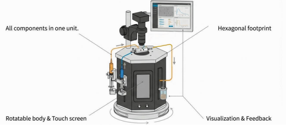
Figure 1. Porous poly(MMA-HEMA) microparticle production station
This workstation combines intuitive syringe pump control with visualization system. It is an integrated solution for any lab wanting to adopt droplet microfluidics technology.
The MONO chip is an assembled glass capillary microfluidic device for single droplet generation. It is composed of fully removable parts: a hexagonal prism-shaped glass chip body with mounting holes, coaxially-aligned capillary tubes and capillary tube adjustment assemblies.
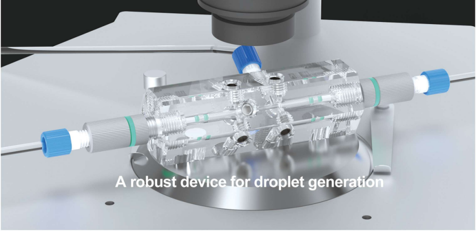
Figure 2. MONO chip design
The primary emulsion is used as the dispersed phase, while a solution of 2% F127, 20% glycerol, and water serves as the continuous phase. Connect syringes containing the primary emulsion (dispersed phase) and continuous phase to the chip inlets via tubing.
Start continous phase first at a low rate to wet channels. Then introduce dispersed phase. Continue both flows until all air bubbles are purged and steady flow is achieved. Adjust the flow rate ratio (continous phase:dispersed phase) to tune droplet size. Normally, higher flow rate ratio = Smaller droplets.
Free-radical polymerization of the oil phase of (W1/O)/W2 emulsions was initiated by UV irradiation for 12 min in an ice-water bath, which was followed by the conversion of the emulsions into poly(MMA-HEMA) microspheres embedded with numerous water droplets.
The macroporous structures inside poly(MMA-HEMA) microspheres were obtained by removing the water droplets. The resultant macroporous poly(MMA-HEMA) microspheres were washed by ethanol for at least three times to ensure the removal of all unreacted chemicals and surfactants, followed by rinsing with DI water for at least three more times. A series of poly(MMA-HEMA) microspheres were prepared in this way, with volume ratios of water to oil (the oil phase refers to the mixture of HEMA and MMA) being 0%, 20%, and 50%, which are referred to as P-0, P-2, and P-5 microspheres.
The W1/O primary emulsions prepared by the homogeneous emulsification method are fine water droplets dispersed in the oil phase, as shown in Figure 3.
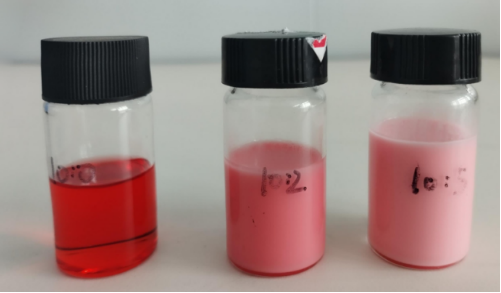
Figure 3. The photographs of W1/O primary emulsions at oil-to-water volume ratios of 10:0, 10:2, and 10:5.
Figure 4 shows the optical microscope images of emulsions and microspheres samples at the room temperature. All emulsions and microspheres with different porogen contents have uniform size, good sphericity and monodispersity. The emulsions without the addition of porogens are transparent (Fig 4(a)). With the addition of W1/O primary emulsions, the transmittance of (W1/O)/W2 emulsions decreases.
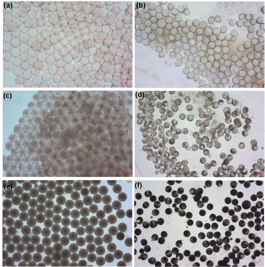
Figure 4. Optical micrographs of (W1/O)/W2 emulsions and poly(MMA-HEMA) microspheres with different volume ratios of oil to water. The volume ratio of oil to water is 10:0 (a, b), 10:2(c,d), 10:5 (e,f).
When the content of porogens is 0% (v/v), the microspheres have a dense structure and there are few pores on the surface of the microspheres (Figure 5(a) and (b)). As the volume ratios of oil to water increases to 20% (v/v), microscale pores begin to appear on the surface (Figure 5(c) and (d)). When the porogens content reaches 50% (v/v), there are a great deal of pore structures in the outer surface (Figure 5(e) and (f)) .
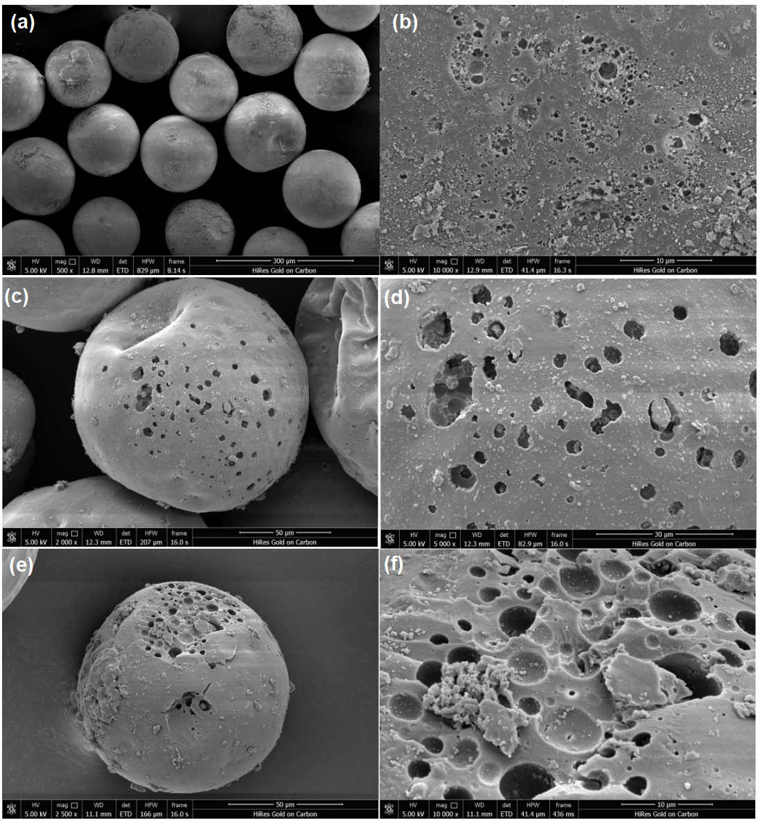
Figure 5. SEM images of poly(MMA-HEMA) microspheres. The P-0, P-2 and P-5 microspheres are shown in the first (a,b), the second (c,d) and third (e,f) row, respectively. Within each row are images of the outer surface, the outer surface with magnification of each type of microspheres.
This study successfully demonstrates the synthesis of monodisperse porous poly(MMA-HEMA) microspheres using a water-in-oil primary emulsion as a template-based foaming agent. SEM analysis confirms that the internal porous microstructure is highly dependent on the foaming agent concentration; as the water content in the primary emulsion increases, both the number and diameter of the pores increase.
B, Ya Lan Yu A , et al. "Monodisperse macroporous microspheres prepared by microfluidic methods and their oil adsorption performance - ScienceDirect." Colloids and Surfaces A: Physicochemical and Engineering Aspects 579.C:123617-123617.