
Mesoporous silica materials have been synthesized for over a decade, often utilizing methods such as evaporation-induced self-assembly (EISA). While EISA can produce ordered structures, the resulting particles typically suffer from a wide size distribution. Monodisperse mesoporous silica microspheres (MMSMs) are of significant value in specialized fields including controlled drug release, biomolecule encapsulation, catalyst preparation, and biochemical sensor design.
Traditional methods like the Stöber process struggle to produce microspheres larger than ten microns. Microfluidic technology breaks this size restriction by precisely controlling droplet formation to create highly uniform liquid templates. The microfluidic-assisted sol–gel preparation of monodisperse silica microspheres primarily consists of three steps: silica sol preparation, monodisperse droplet generation, and the gelation process/high-temperature calcination treatment.
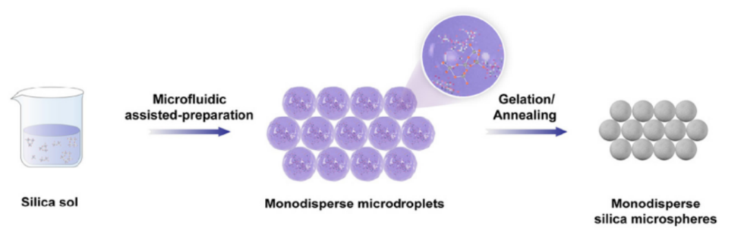
Figure 1. Microfluidic-assisted sol-gel preparation of silica microspheres1.
The experiment utilizes the MF-2G Droplet Microfluidics Workstation equipped with a MONO model capillary microfluidic chip. Monitoring is performed via a high-speed camera and an inverted microscope.
The MONO chip is an assembled glass capillary microfluidic device for single droplet generation. It is composed of fully removable parts: a hexagonal prism-shaped glass chip body with mounting holes, coaxially-aligned capillary tubes and capillary tube adjustment assemblies.
The MONO specific design allows for single emulsions fabrication.
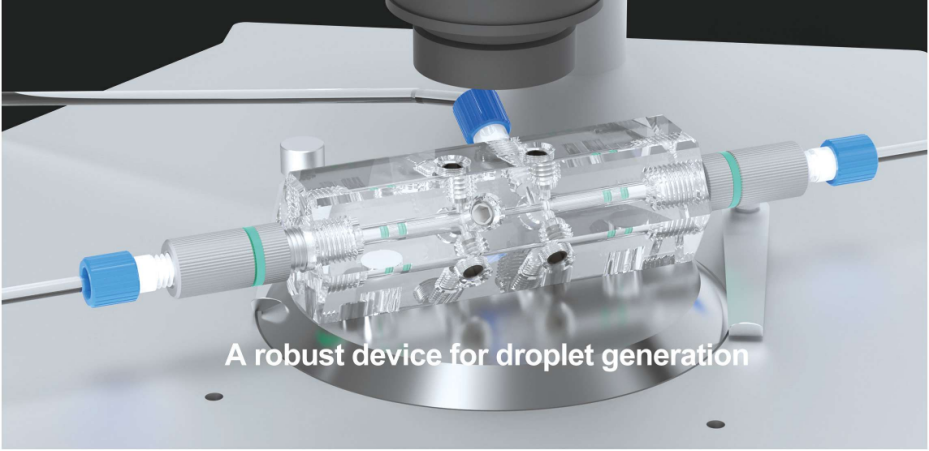
Figure 2. MONO chip design
Hydrolyze TEOS under acidic conditions to form a stable silica sol with low viscosity.
○ Use the MONO chip with nozzle of 70 µm in injection capillary and of 150 µm in collection capillary.
○ Set the dispersed phase flow rate to 5–7 μL/min and the continuous phase flow rate to 100 μL/min.
○ The system generates stable droplets with initial diameters between 35 and 85 μm.
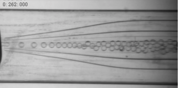
Figure 3. Polystyrene beads produced in MONO chip, the scale is 200 μm.
○ Place the collected droplets in a 70°C oven for 24 hours to allow TEOS to undergo hydrolysis and condensation, forming silica gel microspheres.
○ Centrifuge the mixture with ethyl acetate to remove oil, then wash with ethanol.
○ Dry the particles in a 70°C oven overnight.
Heat the spheres from room temperature to 800°C at a rate of 5°C/min and maintain for 2 hours. This step removes organic residuals and enhances mechanical strength.
The as-formed microdroplets were further dispersed in hexadecane oil, promoting silica gelation and solvent diffusion.The solvent (ethanol and water) in the droplets gradually diffused into the oil phase and evaporated into the air; meanwhile, the silica sol gelation occurred and formed solid microspheres. As shown in Figure 4, the droplet shrinkage was clearly observed. The SEM images of the final microspheres after annealing are presented in Figure 4(c). All the microspheres were in spherical shape with small size distributions.
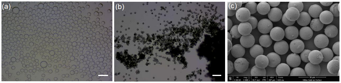
Figure 4. (a) Optical microscopy images of microdroplets. (b) Optical microscopy images of gelated microspheres. (c) SEM image of annealed microspheres.Scale bar: 200 μm.
X-ray diffraction shows a single diffuse peak at approximately 22° (2θ), confirming an amorphous (non-crystalline) structure typical for silica processed below 1700°C.
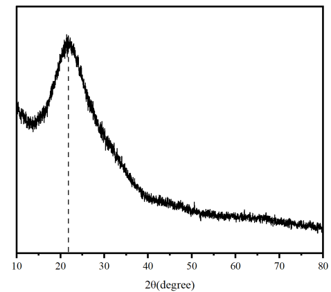
Figure 5. XRD of silica microspheres annealed at 800 °C for 2 h.
The microfluidic-assisted sol–gel strategy successfully produces monodisperse silica microspheres with properties "on demand". This workstation-based solution provides professional precision in controlling particle size and internal mesoporous structures without the need for organic templates. These microspheres are ideal for high-precision applications in chromatography, drug delivery, and advanced sensors.
1) Dai, Zhang , et al. "Microfluidic-assisted sol–gel preparation of monodisperse mesoporous silica microspheres with controlled size, surface morphology, porosity and stiffness." Nanoscale 17.9(2025).
2) Carroll, Nick J. , et al. "Droplet-based microfluidics for emulsion and solvent evaporation synthesis of monodisperse mesoporous silica microspheres. " Langmuir 24.3(2008):658-661.